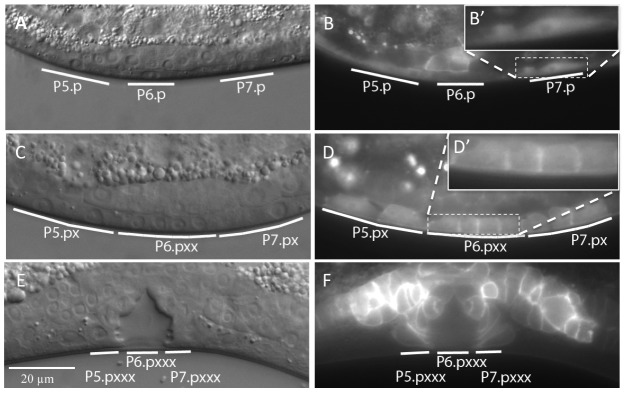
Figure 3. Expression pattern and sub-cellular localization of DAF-18::GFP.
(A and B) Nomarski and fluorescence images of animals expressing the DAF-18::GFP translational reporter at the Pn.p cell stage, (C and D) the Pn.px(x) stage and (E and F) the Pn.pxxx “Christmas tree” stage. The insets in (B′) and (D′) show higher magnifications of the areas in (B) and (D) marked with dashed boxes.

