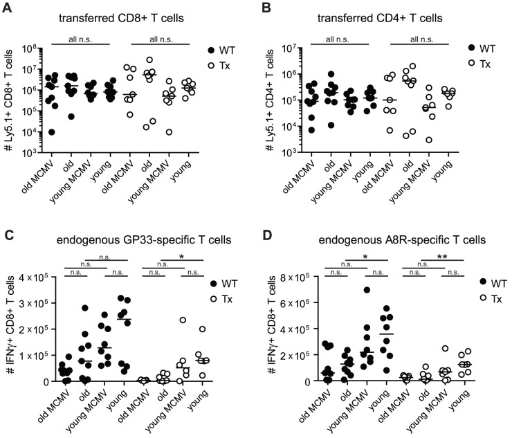
Figure 4. Analysis of virus specific T cell responses after adoptive transfer of TCR transgenic CD8+ and CD4+ T cells and VACV-GP infection in young and old mice with/without Tx or latent MCMV-infection.
Transgenic GP33-specific CD8+ and GP61-specific CD4+ T cells were isolated from the spleen of young Ly5.1+ donor mice and transferred into young (4–6 months old) or old (23–28 months old), MCMV-infected (2–4 or 20–25 months p.i.) or uninfected, Tx (open circles) or non-Tx (wt, closed circles) recipients (Ly5.2). One day later, mice were infected with 5×10⁶ pfu VACV-GP i.p.. Six days later, VACV-GP-specific CD4+ and CD8+ T cell responses were measured in the lung by tetramer-staining (A) or by ICS after in vitro re-stimulation with the respective peptide (B–D) and analysed by flow cytrometry. (A) Total number of GP33-specific transgenic CD8+ T cells (Ly5.1) in the lung. (B) Total number of GP61-specific transgenic CD4+ T cells (Ly5.1) in the lung. (C) Total number of endogenous GP33-specific CD8+ T cells (Ly5.2) in the lung. (D) Total number of endogenous A8R-specific CD8+ T cells in the lung. Pooled data from two independent experiments are displayed. Circles indicate values of individual mice, horizontal bars indicate the medians. Significance was assessed by ANOVA followed by Bonferroni post-analysis (* p<0.05; ** p<0.01; ns = not significant).

