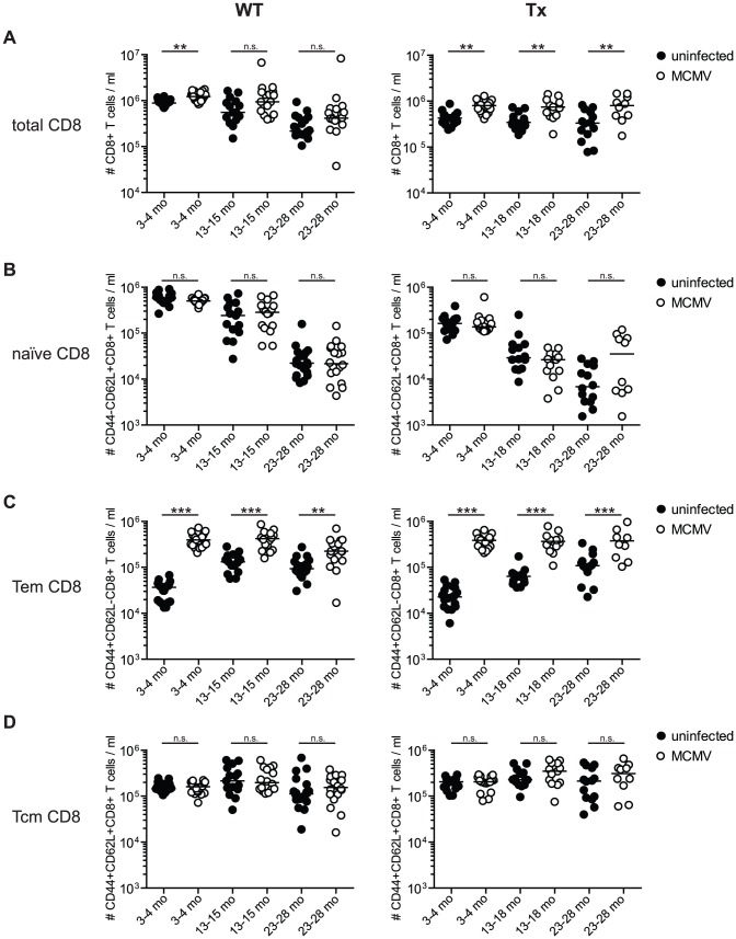
Figure 5. Long-term analysis of the CD8+ T cell compartment after MCMV infection and/or thymectomy in young, middle-aged and old mice.
C57BL/6 mice were thymectomised at age of 4–5 weeks. Two to 3 weeks later, Tx (right panel) and non-Tx wild type (WT, left panel) mice were infected with 107 pfu MCMV-Δ157 i.v. The absolute number of CD8+ T cells was quantified in the blood and phenotypically characterised by polychromatic flow cytometry using CD44 and CD62L. Quantitative analysis of total CD8+ T cell numbers (A), naïve CD8+ T cell numbers (B; CD8+CD44−CD62L+), effector memory (Tem) CD8+ T cell numbers (C; CD8+CD44+CD62L−) and central memory (Tcm) CD8+ T cell numbers (D; CD8+CD44+CD62L+). Data were pooled from three independent experiments. Circles indicate values of individual mice, horizontal bars correspond to the median of an experimental group. Significance was assessed by ANOVA followed by Bonferroni post-analysis (* p<0.05; ** p<0.01; *** p<0.001; ns = not significant).

