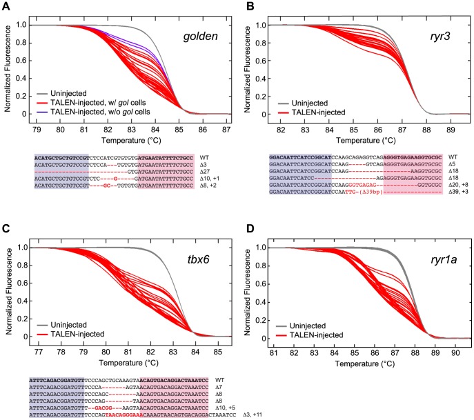
Figure 3. Induction of somatic mutations with TALENs.
(A–D, Upper panels) HRMA detection of targeted mutations in WT embryos that had been injected at the 1 cell stage with RNAs encoding TALENs directed against the golden (A), ryr3 (B), tbx6 (C), or ryr1a (D) gene (see Figure S1 for target sites). gDNA was isolated from individual uninjected or TALEN RNA-injected 1–2 dpf embryos and subjected to HRMA. Each curve is the melting profile of re-annealed amplicons generated from a single embryo. LightScanner Call-IT Software (Idaho Technology) was used to identify melt curves that differed significantly from WT. The results shown here reflect a single experiment, but results from all injections are tablulated in Table 2. Newly induced DNA polymorphisms at the targeted loci were evident in all but one injected embryo (red curves, Table 2), and even gol-ex2 TALEN RNA-injected embryos that did not exhibit patches of pigmentless tissue had induced golden mutations as detected by HRMA (A, purple curves). (A–C, Lower panels) TALEN-induced sequence alterations at targeted loci. Genomic sequences bordering the targeted loci were amplified from embryonic gDNA samples, cloned, and sequenced. Examples of recovered alleles (purple and pink boxed sequences indicate Left and Right RVD binding sites of the TALENs, respectively; red indicates sequence alterations) indicate that insertion/deletion (indel) mutations centered at the TALEN target sites had been induced in somatic tissues of embryos.

