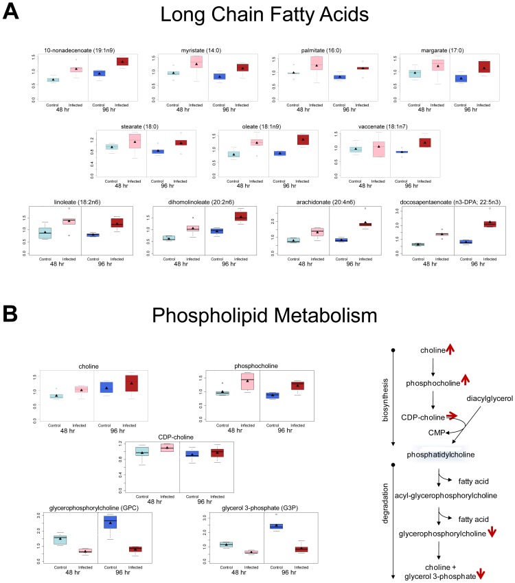
Figure 1. KSHV infection of endothelial cells induces fatty acid production through de novo synthesis.
Relative levels of metabolites from mock- (control) and KSHV-infected (infected) TIME cells are shown at 48 hpi (light blue and pink respectively) and 96 hpi (dark blue and red respectively). A) Box and whisker plots showing relative levels of fatty acid metabolites significantly altered by KSHV infection. B) Box and whisker plots of metabolites that differentiate production of long chain fatty acids (LCFAs) by synthesis or degradation of phospholipids indicating that increased fatty acid metabolites in KSHV infected cells come from increased synthesis.

