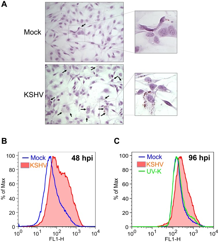
Figure 2. KSHV-infected cells induce the formation of lipid droplet organelles.
A) 48 hpi, TIME cells were fixed and stained with Oil-Red-O, a specific stain for lipid droplets, and hematoxylin, to stain cell nuclei. Cells were visualized by bright field microscopy. B) Mock- and KSHV- infected TIME cells were harvested at 48 hpi, fixed and stained with Nile Red, a specific fluorescent stain for lipid droplets. Staining was analyzed by flow cytometry. C) Mock-, KSHV- and UV-irradiated-KSHV- infected TIME cells were harvested at 96 hpi and stained as in B for lipid droplets.

