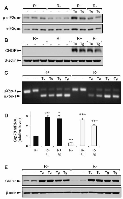
Fig. 4.
IGF-1R knockout does not affect UPR signaling. R+ cells and R− cells were cultured in growth medium containing 4.5 g/L glucose and 10% FBS alone, or growth medium containing ER stress inducing agents tunicamycin (Tu, 1.5 μg/ml) or thapsigargin (Tg, 300nM) for 16 h. A,B: Representative immunoblots for eIF2α phosphorylation and CHOP protein expression. C: Detection of spliced Xbp-1 mRNA by RT-PCR. D: Expression levels for Grp78 mRNA using quantitative real time PCR. E: Representative immunoblot for GRP78 protein induction by Tu and Tg. Values are presented as mean ± SE, n=2-4 *p≤0.05, ***p≤0.001 compared to untreated R+ group. +++p≤0.001 compared to untreated R− group.

