Fig. 6.
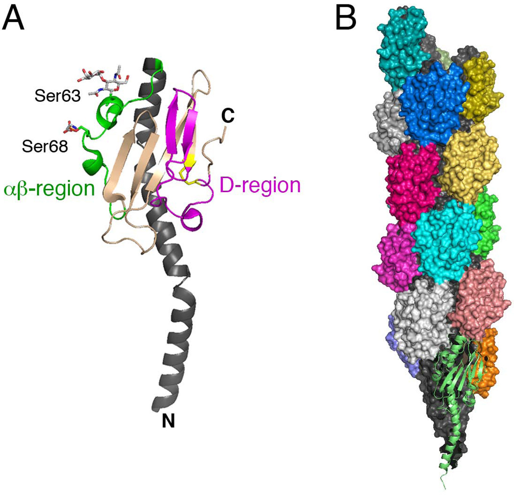
Structures of the N. gonorrhoeae pilin and assembled T4P fiber. (A) Crystal structure of PilE (PDB ID: 2HI2; (Craig et al., 2006)). The N-terminal α-helix is shown in dark gray, the αβ-region in green, and the D-region in magenta with the conserved disulfide bond in yellow. The phosphoethanolamine and disaccharide modifications to serine-68 and -63, respectively, in the αβ-region are shown in stick representation. (B) Atomic model of the T4P fiber based on docking of the PilE structure into a cryoEM density map (PDB ID: 2HIL; (Craig et al., 2006)). The pilin subunits are shown in surface representation, with their N-terminal α-helices in dark gray and surface-exposed C-terminal globular domains colored according to the individual pilin subunits. The bottom-most pilin is depicted in ribbon representation in light green. Images were generated using PyMOL.

