
Fig. 1.
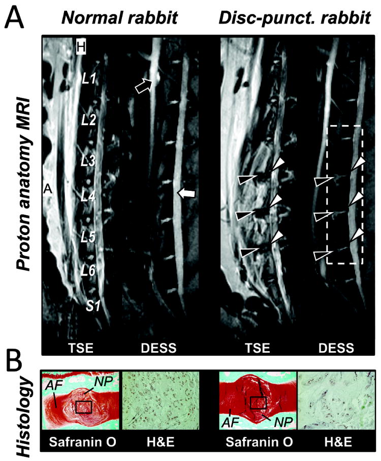
Rabbit lumbar spine MR anatomy and histology. A, 1H MR image of L1 to S1 of the normal and the disc-punctured rabbit; (Left panel) T2-weighted TSE and (right panel) DESS image in sagittal view. Black arrows indicate the descending/ascending aorta vessel and white arrows indicate the CSF in spinal cord. Edema regions (black arrowheads) and degenerated discs (white arrowheads) due to the disc-stab injury. B, Histology images. Safranin O (left panels) and H&E histology images (right panels) acquired from the normal and the injured degenerated discs. Black rectangles are the regions of H&E images. MR parameters were measured in the area indicated by white dotted rectangle in right panel. Abbreviation; A – Anterior and H – head. Same abbreviation was used in the following figures.