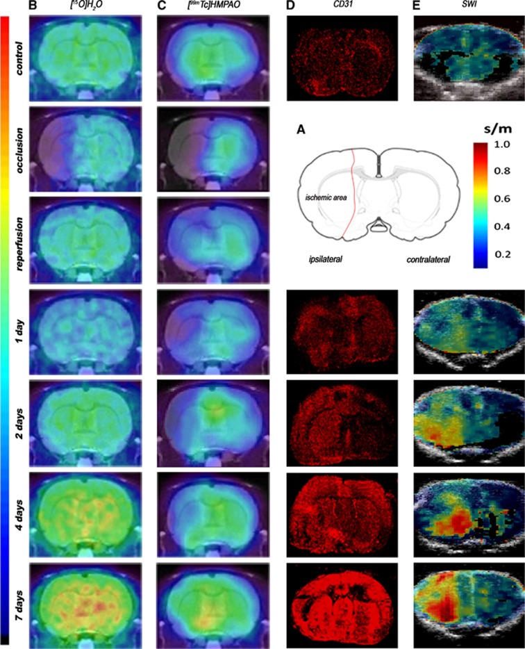
Figure 1.
Serial images of [15O]H2O PET, [99mTc]HMPAO SPECT, CD31 immunohistochemistry, and shear wave imaging (SWI) during occlusion, early reperfusion and at day 1, day 2, day 4, and day 7 after middle cerebral artery occlusion (MCAO). (A) Rat brain section showing a representative ischemic area after MCAO. (B) PET, (C) SPECT, (D) CD31, and (E) SWI images. Panels B and C are coregistered with a magnetic resonance imaging (MRI) (T1) rat template; panel E is coregistered with B-mode images. PET, positron emission tomography; SPECT, single photon emission computed tomography; [99mTc]HMPAO, [99mTc]hexamethylpropylene-amino-oxime.

