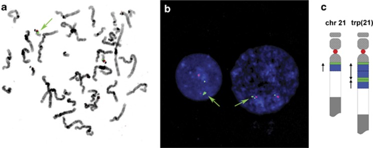
Figure 3.
(a) Metaphase spread and (b) interphase nuclei, counterstained with DAPI, after hybridization with RP11-958H20 (green) and p190.22 (red, specific for the centromeric region of chromosome 22). Green arrows show a larger signal for RP11-958H20 in the metaphase chromosomes and a split signal in the interphase nuclei, in which the signal is split in a larger and a smaller signal. (c) Schematic representation of the proposed arrangement of the triplication in chromosome 22 with in red and green the location of the FISH probes used in panels (a, b).

