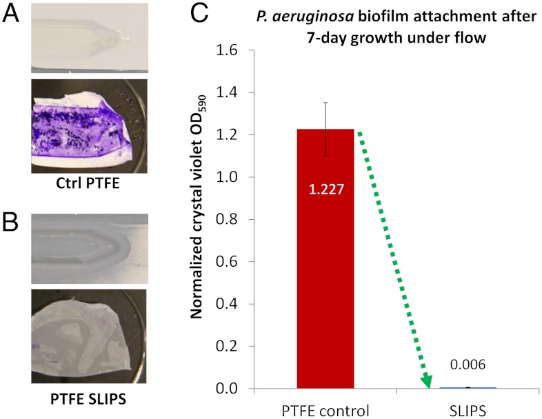
Fig. 3.
Characterization of biofilm attachment to SLIPS and PTFE in a dual-chamber flow cell with 10 mL/ min flow rate. (A–B) Photographs of the control PTFE and SLIPS substrates after the flow cell was opened following 48 h growth, both before (Top) and after (Bottom) crystal violet staining. (C) Crystal violet staining-based quantification of accumulated biomass on SLIPS versus control PTFE following 7 d of growth.

