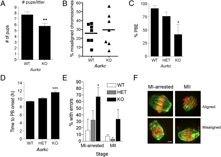
Fig. 1.
Loss of AURKC leads to meiotic abnormalities. (A) Results of fertility trials. The average number of pups born to females of the indicated genotype over an 8-mo breeding trial is shown. (B) GV-intact oocytes were isolated from mice of the indicated genotype and matured in vitro for 8 h before fixation at MI. Percent of oocytes with misaligned chromosomes was plotted for each mouse analyzed. (C–F) GV-intact oocytes were isolated from mice of the indicated genotype and matured in vitro to determine incidence (B) and timing (C) of polar body extrusion (PBE). Cells were fixed when controls (WT) had reached MII and processed for immunocytochemistry to detect chromosomes and spindles. The percentage of oocytes that contained abnormal chromosome configurations at either MI or MII was determined (E), and representative images are shown (F). Graphs represent mean (± SEM) from at least 30 oocytes from three independent experiments. (Scale bars, 5 μm.) One-way ANOVA was used to analyze the data in B–D. *P < 0.05, **P < 0.01, ***P < 0.001; WT, wild-type; HET, heterozygous; KO, knockout.

