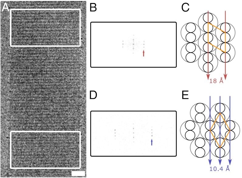
Fig. 2.
Direct observation of ultrastructure in frozen-hydrated SAFs. (A) Representative cryo-TEM of a single frozen-hydrated SAF, revealing the lateral striations across the width and along the length of the entire fiber (fiber axis is vertical), and two different sets of longitudinal striations (boxed regions). Scale bar, 20 nm. (B) Fourier transform of A, Upper, with the first row line at 18 Å (arrow). (C) Projecting along the [100] axis (arrows) causes alignment of the 18 Å [100] planes of the unit cell. Small circles represent α-helices within the coiled coil (large gray circles); the hexagonal unit cell with sides of 20.8 Å is shown in orange. (D) Fourier transform of A, Lower, with the row line at 10.4 Å (arrow). In both B and D, the first meridional reflection is at 41.8 Å. (E) Projecting along the [110] axis aligns the 10.4 Å [110] planes.

