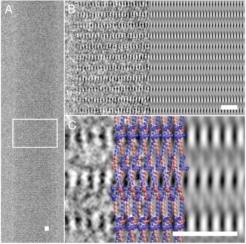
Fig. 3.
Direct imaging of dimeric α-helical coiled coils within the SAF ultrastructure. (A) Region showing [100] planes to high resolution used for further analysis (fiber axis is vertical). (B) After processing the images with 2dx (28), the magnified boxed area (Left) more clearly shows the regular lattice of high-contrast striations (Right). (C) Further magnification of the experimental (Left) and processed (Right) images, with a cartoon overlay of the postulated SAF structure (Middle, colored as in Fig. 1). Light areas indicate high electron density. The lateral striations are proposed to be caused by the charged Arg and Asp residues (spheres) forming salt bridges between adjacent coiled coils. Scale bars, 10 nm.

