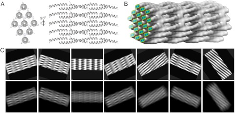
Fig. 5.
Model of the SAF superstructure used to simulate cryo-TEM images. (A) The hexagonal array of coiled coils used for the molecular-dynamics simulations, shown as a sticky-ended unit cell, held together with Asp:Arg salt bridges. (B) Model filtered to 10 Å used to simulate projection images, which is identical to the molecular-dynamics simulation but contains more coiled coils. The surface threshold is set to contain 100% of the mass of the protein array. Protein occupies 50% of the volume of the array and contains pores of approximately 100 Å2. The colored plane shows the density gradient and clearly shows the two α-helices comprising the coiled coils. (C) Projection images of the model generated by EMAN2 (16) (Bottom) and matching 2dx-processed images (28) (Top).

