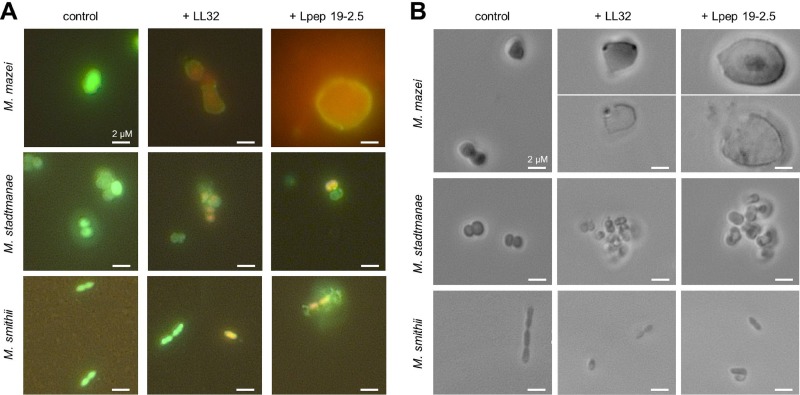
Fig 3.
Microscopic analysis of methanoarchaea after treatment with LL32 and Lpep 19-2.5. Cultures of M. mazei, M. stadtmanae, and M. smithii were grown to the mid-exponential growth phase, LIVE/DEAD staining kit reagents were added according to the manufacturer's protocol, and 1-ml aliquots of cultures were dispensed to anaerobic Hungate tubes. AMPs were added at the following concentrations: M. mazei, 1 μM LL32 or 1 μM Lpep 19-2.5; M. stadtmanae, 5 μM LL32 or 10 μM Lpep 19-2.5; M. smithii, 1 μM LL32 or 3 μM Lpep 19-2.5. (A) Fluorescent micrographs with filter sets for propidium iodide and SYTO 9 fluorescence taken after 10 min of incubation in the presence of AMPs (or in the absence for control cultures). (B) Phase-contrast micrographs taken after 60 min of incubation in the presence of AMPs (or in the absence for control cultures). Pictures are representative for the respective sample.

