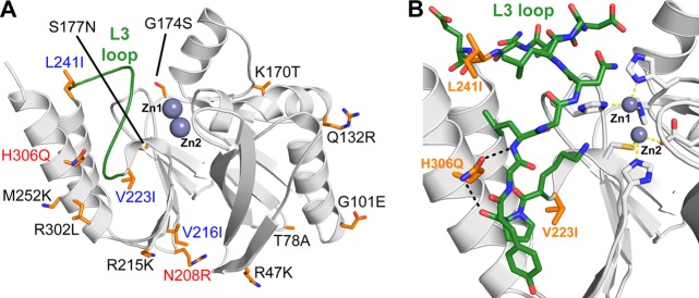
Fig 2.
(A) Molecular representation of the structure of IMP-1 (Protein Data Bank code 1DDK). The 15 residues that are different in IMP-28 are shown as orange sticks, surface residues are black, internal hydrophobic residues are blue, and polar internal residues are red. (B) Enlarged view of the L3 loop of IMP-1 (green).

