
Fig 3.
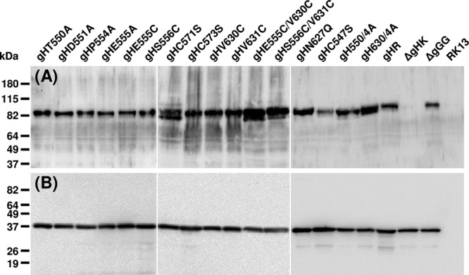
Western blot analyses. RK13 cells were harvested 20 h after infection with pPrV-ΔgGG, pPrV-ΔgGGΔgH K, or virus rescuants expressing the indicated gH mutants at an MOI of 5. Proteins were separated by SDS-PAGE and blots were analyzed with monospecific antisera against gH (A) or capsid protein pUL38 (B). Molecular mass markers are indicated on the left.