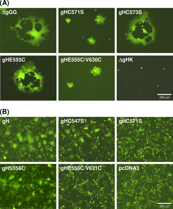
Fig 4.

EGFP autofluorescence illustrating gH function. (A) Cell-to-cell spread was analyzed 1 day after infection of RK13 cells with pPrV-ΔgGG, gH deletion mutant pPrV-ΔgGGΔgHK, or virus recombinants possessing the indicated gH mutations (gHC571S, gHC573S, gHE555C, gHE555C/V630C). (B) Induction of syncytium formation was analyzed 3 days after cotransfection of RK13 cells with an EGFP reporter plasmid and expression plasmids for PrV gB, gD, and gL, as well as for wild-type (pcDNA-gH) or mutated gH (gHC547S, gHC571S, gHS556C, gHS556C/V631C) or the empty expression vector (pcDNA3).
