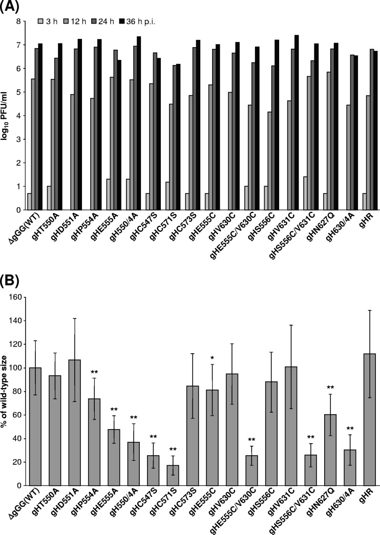
Fig 5.
In vitro replication of PrV gH mutants. (A) For analysis of one-step growth kinetics, RK13 cells were infected at an MOI of 5 and harvested together with the supernatant after 3 h, 12 h, 24 h, or 36 h. Mean progeny virus titers of two experiments are shown. (B) For determination of plaque sizes, infected RK13 cells were incubated for 2 days under semisolid medium. Mean areas of 30 plaques per virus were calculated as percentages of wild-type (pPrV-ΔgGG) sizes. Standard deviations (vertical lines) and statistically significant differences from wild-type size are indicated (*, P < 0.05; **, P < 0.01).

