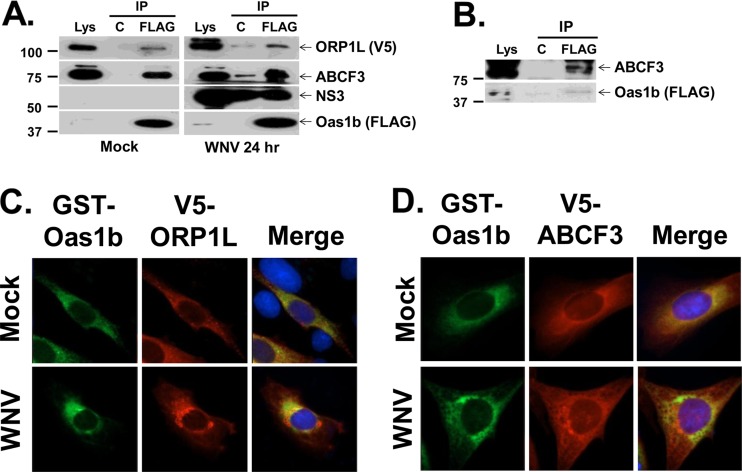
Fig 3.
Coimmunoprecipitation of ORP1L and ABCF3 with Oas1b from mammalian cell lysates. (A) He-Oas1b/ORP1L MEFs that stably express FLAG-tagged Oas1b and V5-tagged ORP1L were either mock infected or infected with WNV Eg101 (MOI of 1), cross-linked with DSP, quenched, and lysed at 24 h after infection. Lysates were immunoprecipitated with either a control antibody or anti-FLAG antibody, and immunoprecipitated (IP) proteins were detected by Western blotting: ORP1L (anti-V5 antibody), ABCF3 (anti-ABCF3 antibody), NS3 (anti-NS3 antibody), and Oas1b protein (anti-FLAG antibody). Each experiment was repeated at least twice, and a representative blot is shown. Lys, lysate; C, control (anti-IgG antibody-conjugated agarose); FLAG, anti-FLAG antibody-conjugated agarose. The positions of protein size markers are indicated on the left. (B) Cell lysates from He-Oas1b cells that stably express FLAG-tagged Oas1b were immunoprecipitated with either a control antibody or anti-FLAG antibody, and immunoprecipitated proteins were detected by Western blotting. Plasmids expressing GST-Oas1b and V5-ORP1L (C) or V5-ABCF3 (D) were transiently cotransfected into BHK cells, and 24 h later cells were either mock infected or infected with WNV Eg101 (MOI of 5). At 24 h after infection, cells were fixed, permeabilized, and analyzed by immunofluorescence. Anti-GST antibody (green) detected GST-Oas1b, anti-V5 antibody (red) detected V5-tagged proteins, and Hoechst stain (blue) detected nuclei.

