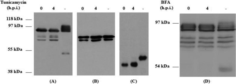
Fig 2.
Western blot analysis of the glycosylation patterns of HEV recombinant rORF2-vv (A and D) and rΔ(1-111)-ORF2-vv (B) proteins and of B5 WR vaccinia virus protein (C) in infected BHK-21 cells. Western blots were stained with a monoclonal anti-His antibody (A, B, and D) or with an anti-B5 rat monoclonal antibody (C). Infected-cell extracts treated with the drugs at 0 or 4 h p.i. or not treated (−) were harvested 7 h p.i. and processed as described in Materials and Methods. Molecular mass markers are shown on the left.

