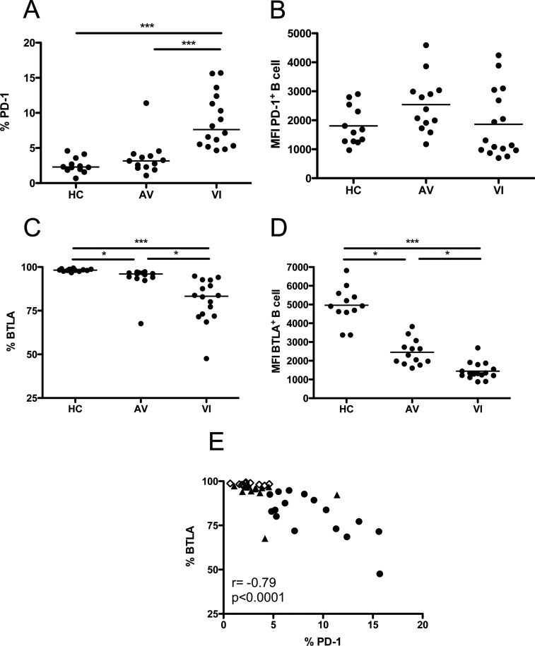
Fig 1.
Expression of PD-1 and BTLA by B lymphocytes. (A and C) Percentages of total B cells (CD19+) that express PD-1 (A) and BTLA (C) in HC, AV, and VI subjects. (B and D) Mean fluorescence intensity (MFI) for PD-1 (B) and BTLA (D) expression by individual PD-1+ or BTLA+ CD19+ B cells. Each point represents data from a single subject. Horizontal bars within the point plots indicate the median percentage for each group. Significance between groups determined by 1-way ANOVA is indicated above the groups: *, P < 0.05; **, P < 0.01; ***, P < 0.001. (E) Correlation between percentages of total B cells that express PD-1 and BTLA. Spearman's rank correlation coefficient (r) and level of significance (p) are indicated within the graph. Open diamonds, HC; closed triangles, AV; closed circles, VI.

