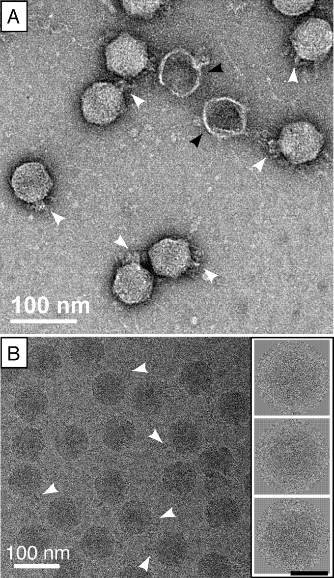
Fig 4.
Electron microscopy of CW02 by negative stain (A) and cryogenic (B) methods. Stubby or thin tail-like features are marked by white arrowheads. Collapsed, empty particles are shown by black arrowheads. The inset in panel B shows examples of particle images used in computing the 3D reconstruction (black scale bar, 50 nm).

