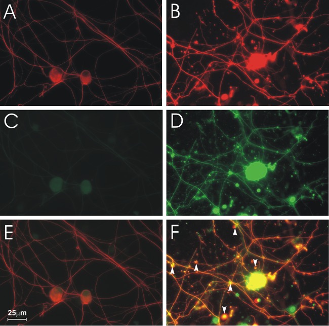
Fig 1.
Formation of axonal swellings containing 4-HNE adducts in DRG cultures. Fluorescence microscopy showing mock-infected (A, C, E) and CVS-infected (B, D, F) cultures. DRG neurons stained for β-tubulin (A, B) showed large axonal swellings in CVS-infected neurons (B). Staining for 4-HNE adducts (C, D) showed that CVS infection induced intense staining for 4-HNE adducts (D) at 72 h postinfection (p.i.) in comparison to mock-infected DRG neurons (C). Merging signals for β-tubulin and 4-HNE (E, F) showed that 4-HNE was expressed in axonal swellings (arrowheads) in CVS infection (yellow) (F).

