
Fig 2.
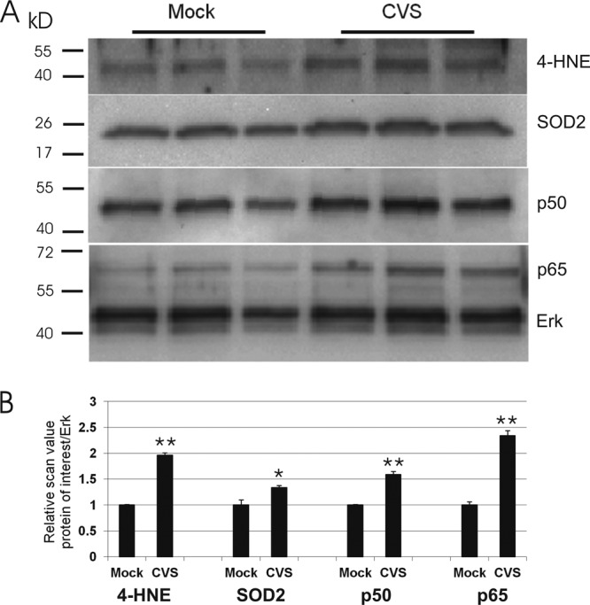
CVS infection altered the protein expression of the p50 and p65 subunits of NF-κB, 4-HNE adducts, and SOD2. Cell protein lysates from mock- and CVS-infected DRG cultures 72 h p.i. were loaded on 10% Tris acrylamide gel. (A) Western blot of p50, p65, 4-HNE, SOD2, and Erk, which served as the total protein loading control. (B) Relative scan values on 3 triplicates show a 1.6-fold increase in p50 (0.75 ± 0.01 versus 1.20 ± 0.06, P = 0.002), 2.4-fold increase in p65 (0.56 ± 0.06 versus 1.34 ± 0.10, P = 0.0022), 2.0-fold increase in 4-HNE (0.65 ± 0.01 versus 1.28 ± 0.05, P = 0.0002), and 1.3-fold increase in SOD2 (0.84 ± 0.11 versus 1.14 ± 0.04, P = 0.047) in CVS infection versus mock infection. Statistical significance is indicated by * (P < 0.05) and ** (P < 0.01).