Fig 1.
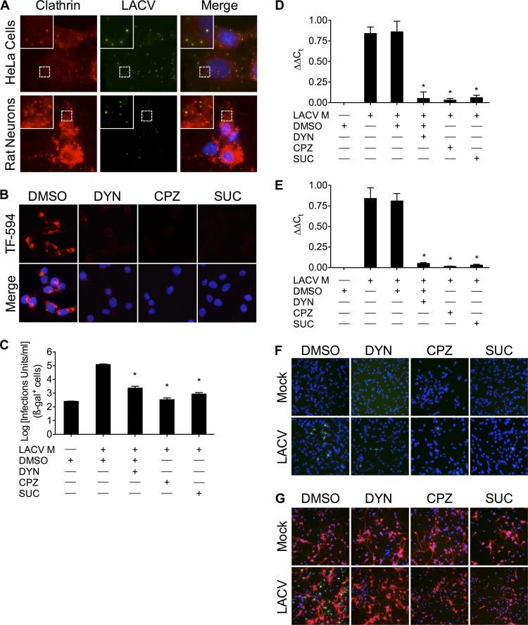
LACV requires dynamin- and clathrin-mediated endocytosis during early infection in HeLa cells and primary neurons. (A) Colocalization of LACV with clathrin shortly after LACV internalization. LACV (MOI, >10) was incubated with either HeLa cells or primary rat neuronal cultures on ice for 90 min. Cells were then slowly shifted to 37°C to allow LACV uptake and fixed at 0 to 20 min after adsorption/during warming (images shown were taken at 20 min after the onset of warming). Cells were then permeabilized and stained with anti-LACV Gc and anti-clathrin antibodies, followed by secondary antibodies conjugated to FITC and R-phycoerythrin, respectively. Images were taken of four z-sections of the same field and then overlaid. Nuclei (blue) were stained with Hoecht's stain. Green, LACV Gc; red, endogenous clathrin. (B) Dynasore (DYN), chlorpromazine (CPZ), and hypertonic medium blocks transferrin uptake. BHK-21 cells (untreated or pretreated with DMSO [0.4%, vol/vol], DYN [40 or 80 μM], CPZ [5 or 10 μg/ml], or sucrose [SUC; 0.3 or 0.45 M]) were incubated with Alexa Fluor-594-conjugated transferrin (TF-594; red). After 45 min, cells were fixed and analyzed by fluorescence microscopy for the uptake of transferrin. (C) MLV pseudotypes of LACV transduce CHME-5 cells less efficiently in the presence of DYN, CPZ, or hypertonic medium treatment (*, P < 0.01 by Tukey test). (D to G) DYN, CPZ, and hypertonic medium inhibit LACV entry in BHK-21 cells and primary rat neurons. Following 30 min of pretreatment with DYN, CPZ, or hypertonic medium, BHK-21 cells (D and F) or primary rat neuronal cultures (E and G) were mock infected or infected with LACV (MOI, 0.5 [C to E] or 0.2 [F and G]) in the presence of inhibitors of clathrin-mediated endocytosis for 1 h. RNA from cell lysates 6 h postinfection was isolated and subjected to qPCR using LACV M segment-specific primers. DYN, CPZ, and hypertonic medium decreased LACV M segment RNA (relative to 18S RNA) compared to LACV-infected, control, or DMSO-treated cells (*, P < 0.001 by Student Newman-Keuls test). Ct, threshold cycle. The results are means ± standard deviations (SD) from 3 independent experiments. BHK-21 cells (F) and primary rat neuronal cultures (G) were fixed 8 h after infection, permeabilized, and stained with anti-LACV Gc (green) and anti-MAP2 (G; red) antibodies. Only higher concentrations of each treatment are shown in panels B to G.

