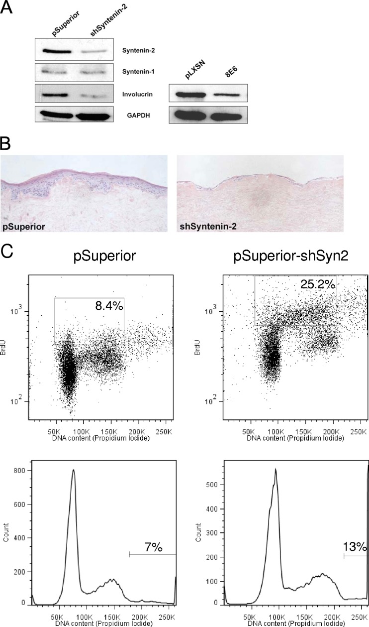
Fig 7.
Specific knockdown of syntenin-2 led to changes in keratinocyte differentiation and proliferation. (A) Western blot analysis of syntenin-2, syntenin-1, and involucrin in keratinocytes infected with the empty retroviral construct pSuperior or retroviral constructs coding for shSyntenin-2. Involucrin levels were also analyzed in keratinocytes expressing pLXSN or HPV8 E6. GAPDH was used as the loading control. (B) Organotypic skin cultures of PHEK expressing the empty retroviral construct pSuperior or the construct coding for shSyntenin-2. These were repeated three times, and sections of representative cultures are shown which were stained with hematoxylin and eosin and analyzed for histological changes. Magnification, ×200. (C) Flow cytometric analysis of pSuperior- and shSyntenin-2-expressing cells. Dot plots (top; gated on cells in the S-phase of the cell cycle), and histogram illustrations (bottom; gated on polyploid cells) of BrdU- and propidium iodide-treated cells.

