
Fig 2.
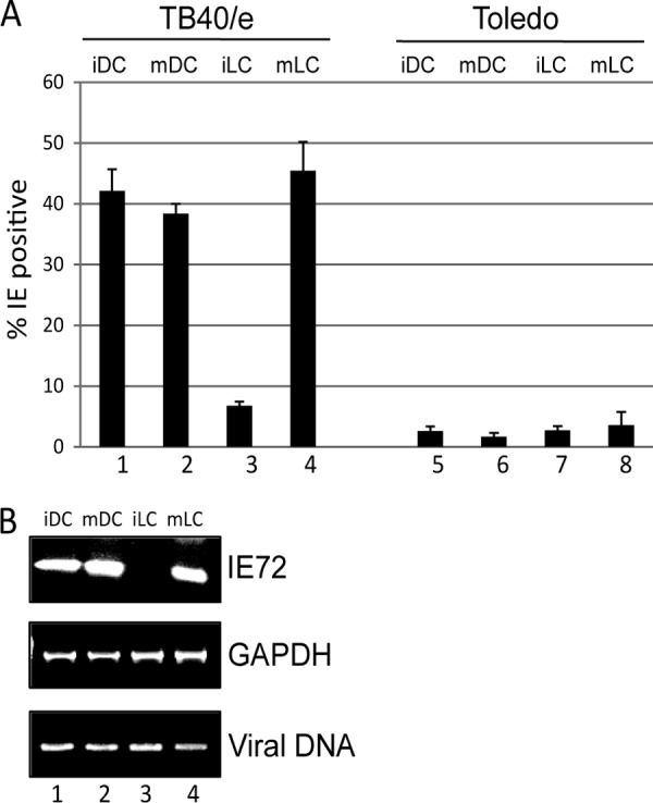
A clinical isolate of HCMV infects immature LCs inefficiently. (A) MoLCs and DCs were infected with TB40/e (columns 1 to 4) or Toledo (columns 5 to 8) before (iDC and iLC) or after (mDC and mLC) LPS stimulation and analyzed for IE gene expression by immunofluorescence microscopy at 24 h postinfection. The percent infection was calculated from four fields per well from an analysis performed in triplicate. (B) MoLCs and DCs were infected with TB40/e before (iDC and iLC) or after (mDC and mLC) LPS stimulation and analyzed by PCR 24 h postinfection for IE72 and GAPDH RNA expression and the presence of viral genomes.