
Fig 5.
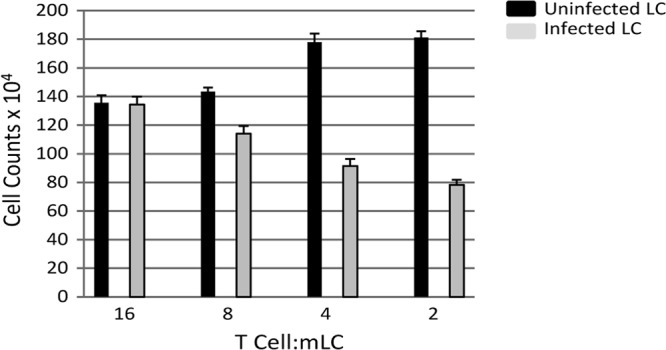
Infected mature MoLCs are impaired in their ability to promote T cell proliferation in a mixed leukocyte reaction. MoLCs were either mock infected (black) or HCMV infected (gray), followed by incubation, at 72 h postinfection, with 8 × 10⁴ T cells at the E:T ratio shown. The T cell proliferation was assayed for the total cell count from triplicate wells. The results shown are representative of four independent repeats.