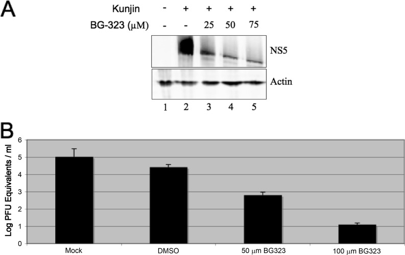
Fig 3.
(A) Western blot analysis of Kunjin virus-infected BHK cells treated with BG-323. BHK cells were infected at an MOI of 0.01 with Kunjin virus and treated with the indicated concentration of BG-323. At 48 h postinfection, cells were collected and NS5 and β-actin proteins were detected by Western blotting. (B) Detection of viral RNA in BG-323-treated medium. BHK cells were infected with Kunjin virus at an MOI of 0.01, and cells were mock treated or treated with DMSO, 50 μM BG-323, or 100 μM BG-323 at the time of infection. Cells were incubated for 48 h, and medium samples were collected and stored at −80°C. Samples were thawed, RNA was extracted, and RNA was quantified by qRT-PCR. qRT-PCR Cq values were compared to those of a Kunjin virus RNA standard curve and converted to log10 PFU equivalents/ml. n = 3.

