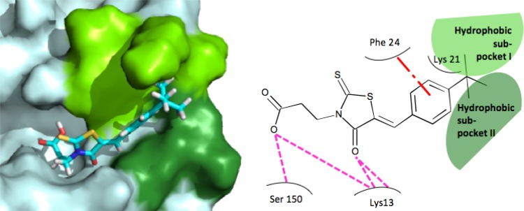
Fig 5.
Molecular docking of BG-323 with the capping enzyme. On the left, a three-dimensional rendering of the predicted orientation of BG-323 within the yellow fever virus capping enzyme is presented. In the yellow fever virus protein, light blue represents the entire protein, bright green represents the location of hydrophobic subpocket 1, and dark green represents the location of hydrophobic subpocket 2. In the stick form depiction of BG-323, carbon atoms are cyan, hydrogen atoms are white, sulfur atoms are yellow, oxygen atoms are red, and nitrogen atoms are blue. On the right, a flatland model of predicted interactions between of BG-323 and the yellow fever virus capping enzyme is presented. BG-323 is depicted in line form surrounded by capping enzyme residues. Light green represents the location of hydrophobic subpocket 1, and dark green represents the location of hydrophobic subpocket 2. The dash-and-dot red line represents the predicted π-π stacking interactions, and the pink dashed lines represent predicted hydrogen bonds.

