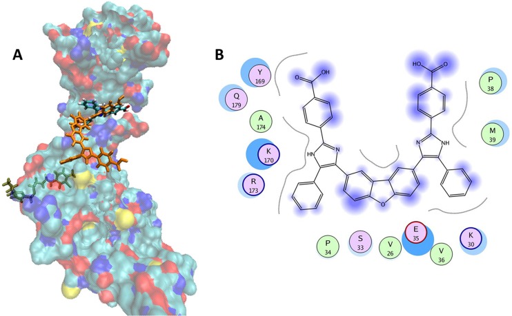
Fig 2.
Proposed binding mode of CK026 to the HIV-1NL4-3 capsid protein. (A) Surface representation of the monomeric unit of CA protein colored by atom type (C in cyan, O in red, N in blue, and S in yellow). CK026, I-XW-053, and a known CA inhibitor, CAP-1, are docked in their predicted binding sites and are shown as licorice models that are orange, colored by atom type, and green, respectively. (B) Proposed binding mode of CK026 in CA. The binding site residues are colored by their nature, with hydrophobic residues in green, polar residues in purple, and charged residues highlighted with bold contours. Blue spheres and contours indicate matching regions between ligand and receptors. The figure was generated using the MOE ligX module.

