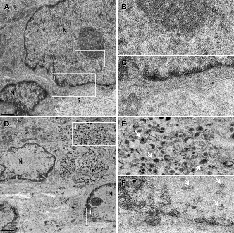
Fig 5.
TEM analysis of infected DRG xenografts shows no evidence of virion production in the 7D samples. TEM images taken from 7D (A to C)- and WT (D to F)-infected DRG samples. The nuclei of sensory neurons (N) and surrounding satellite cells (S) are labeled. In the 7D sample, no evidence of intranuclear capsids (B; box I in panel A) or of cytoplasmic virions (C; box II in panel A) and no tissue damage were observed, whereas the WT-infected DRG sample shows some vacuolization in the plasma membrane region (D), accompanied by the presence of polymorphic virions (white arrows) in the cytoplasm of the infected neuron (E; box I in panel D), as well as intranuclear capsids (F; box II in panel D).

