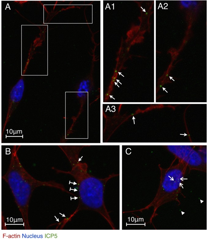
Fig 2.
localization of HSV-1 particles in SK-N-SH neuroblastoma cells during early infection. Cells were infected with HSV-1 (MOI = 10) for 0.5 h and then fixed, permeabilized, and stained with an HSV-1 ICP5 primary antibody, an Alexa Fluor 488-conjugated secondary antibody, TRITC-phalloidin, and DAPI. The images were recorded by LSM. (A) Two infected neuronal cells. (A1 and A2) Higher magnifications of the vertical boxed areas showing viral particles attached to actin-rich dendrites (arrows). (A3) Higher magnification of the horizontal boxed area showing viral particles attached to actin-rich filopodia (arrows). (B) Infected cells with viral particles attached to the surface of the cellular membrane (arrows with tails), the dendritic filopodia (bottom two arrows), and the lamellipodia (the top arrow). (C) Infected cell with viral particles docked at the nucleus (arrows). Free viral particles (arrowheads) are also shown.

