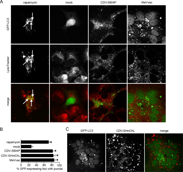
Fig 1.
Morbillivirus infection rapidly induces autophagy. (A) Confocal microscopy analysis of VerodogSLAMtag cells transfected with a GFP-LC3 expression plasmid and infected with CDV 5804P or MeVvac at an MOI of 0.01 or left uninfected. Rapamycin-treated cells were included as an autophagy control. At different times after infection, cells were stained with Lysotracker red, fixed with 4% paraformaldehyde, and imaged by confocal laser microscopy. Results for the 20-h time point are shown. The localization of autophagosomes is indicated by green puncta, and lysosomes are marked in red. Arrows indicate colocalization of GFP-LC3 puncta with lysosomes. (B) Proportion of GFP-expressing cells with punctum formation. For each sample, the total number of GFP-expressing foci and the number of foci with GFP puncta were counted in four separate fields. Data from at least four independent experiments were used for the analysis. Error bars represent the standard deviation, and statistically significant differences (ANOVA, P < 0.05) are indicated by asterisks. (C) Confocal microscopy analysis of GFP-LC3-transfected VerodogSLAMtag cells infected with CDV-SHmChL at an MOI of 0.01. Twenty hours after infection, cells were fixed with 4% paraformaldehyde and visualized by confocal microscopy. The localization of autophagosomes is indicated by green puncta, and that of the CDV polymerase (L)-mCherry fusion protein, indicative of the CDV replication complexes, is marked in red.

