
Fig 4.
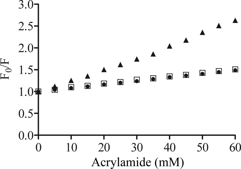
Stern-Volmer plot, showing the acrylamide quenching of the bovicin HC5 tryptophan residue measured for samples containing 1 μM bovicin HC5 in the absence (▲) or presence of DOPC (●) or DLPC/DMoPC (□) vesicles containing 1 mol% of lipid II (100 μM lipid-Pi). Single-wavelength recordings were performed at 340 nm using an excitation wavelength of 280 nm. F0, fluorescence measured in the absence of the quencher; F, fluorescence measured in the presence of the quencher.