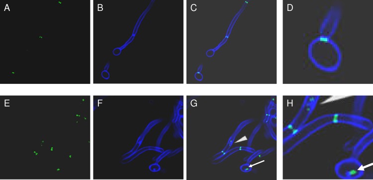
Fig 5.
Septins and chitin colocalize in mutant cells. C. albicans SC5314 and irs4 and inp51 mutants expressing Cdc10-GFP were visualized by confocal microscopy, and chitin was localized by calcofluor white staining. Cdc10-GFP and calcofluor white were distributed normally in SC5314 cells (A and B, respectively), but were concentrated within aberrant patches in the inp51 mutant (E and F, respectively). As evident in the overlay images, Cdc10-GFP colocalized with calcofluor white in the inp51 mutant (G and H [higher resolution], cyan color and arrowheads).

