Figure 1.

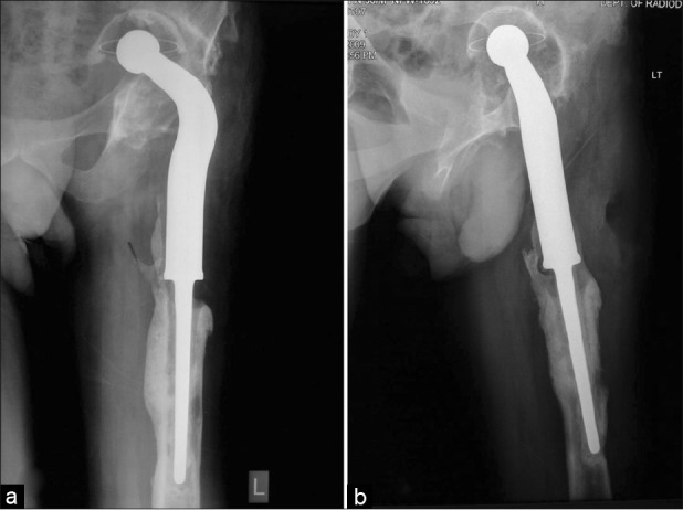
Preoperative radiographs of left hip and thigh anteroposterior (a) and lateral views (b), showing a Paprosky type 3b acetabular defect along with intrapelvic migration of acetabular and femoral component and areas of loosening

Preoperative radiographs of left hip and thigh anteroposterior (a) and lateral views (b), showing a Paprosky type 3b acetabular defect along with intrapelvic migration of acetabular and femoral component and areas of loosening