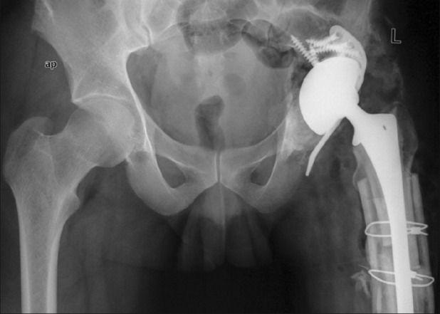
Figure 3.

Immediate postoperative radiograph of pelvis with bilateral hips anteroposterior view following the second stage reconstruction illustrating a well-constructed acetabular and femoral defect with cup-cage construct and allograft prosthesis composite, anatomic restoration of the hip center, and equalization of limb length. The inferior flange of the cage had cut through the ischium
