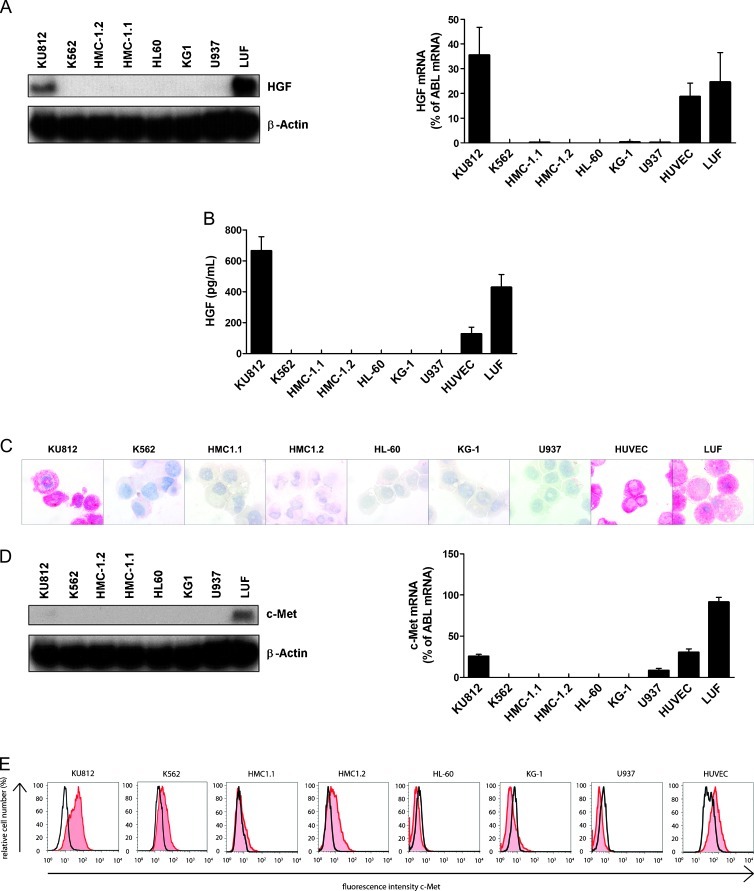
Figure 3.
Expression of HGF and c-Met in various leukemic cell lines. (A) Evaluation of expression of HGF mRNA in various leukemic cell lines, and LUFs by Northern blot analysis (NB left side) and qPCR (right side). β-Actin (NB) and ABL (qPCR) served as control. (B) Measurement of HGF in supernatants of leukemic cell lines, HUVECs, and LUF. Supernatants were obtained after culturing cells in medium with 10% FCS for 5 days. HGF concentrations were determined by ELISA. Results represent the mean ± SD of three independent experiments. (C) ICC evaluation of HGF expression in leukemic cell lines, LUFs and HUVECs. Cells were spun on cytospin slides and stained with an antibody against HGF. Magnification, 100x/1.35. (D) Evaluation of expression of c-Met mRNA in various leukemic cell lines and LUFs by Northern blot analysis (NB; left panel) and qPCR (right panel). β-Actin (NB) and ABL (qPCR) served as control. (E) Surface expression of c-Met on KU812, K562, HMC-1.1, HMC-1.2, HL60, KG-1, U937, LUF, and HUVEC. Cells were analyzed for expression of c-Met by flow cytometry. Expression of c-Met (red histograms) was controlled by an isotype-matched antibody (black open histograms).

