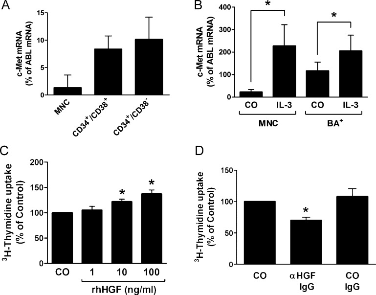
Figure 6.
Expression of c-Met on CML progenitor cells and CML basophils. (A) Expression of c-Met mRNA in highly enriched (sorted) CD34+/CD38- stem cells and CD34+/CD38+ progenitor cells obtained from patients with CML in AP as determined by qPCR. Cells were subjected to RNA isolation, cDNA synthesis, and qPCR using primers specific for c-Met and ABL. Results show c-Met mRNA levels as percent of ABL mRNA levels and represent the mean ± SD of three independent experiments (three patients). (B) Expression of c-Met mRNA in PB MNCs and highly purified CD203c+ basophils (BA+) from three patients with CML. MNCs and basophils were incubated in the absence (CO) or presence of IL-3 (100 ng/ml) for 30 minutes and were then subjected to RNA isolation, cDNA synthesis, and qPCR using primers specific for c-Met and ABL. Results show HGF mRNA expression levels as percent of ABLmRNA levels and represent the mean ± SD of three independent experiments (three donors). *P < .05. (C) KU812 cells were incubated in control medium (CO) or in medium containing various concentrations of rhHGF at 37°C for 48 hours. Then, uptake of 3H-thymidine was measured. Results are expressed as percent of control and represent the mean ± SD of three independent experiments. *P < .05. (D) KU812 cells were incubated in control medium (CO) or in medium containing an anti-HGF antibody (αHGF; 1 µg/ml) or a control antibody (CO IgG; 1 µg/ml) at 37°C for 48 hours. Then, uptake of 3H-thymidine was measured. Results are expressed as percent of CO and represent the mean ± SD of three independent experiments. *P < .05.

