
Fig. 3.
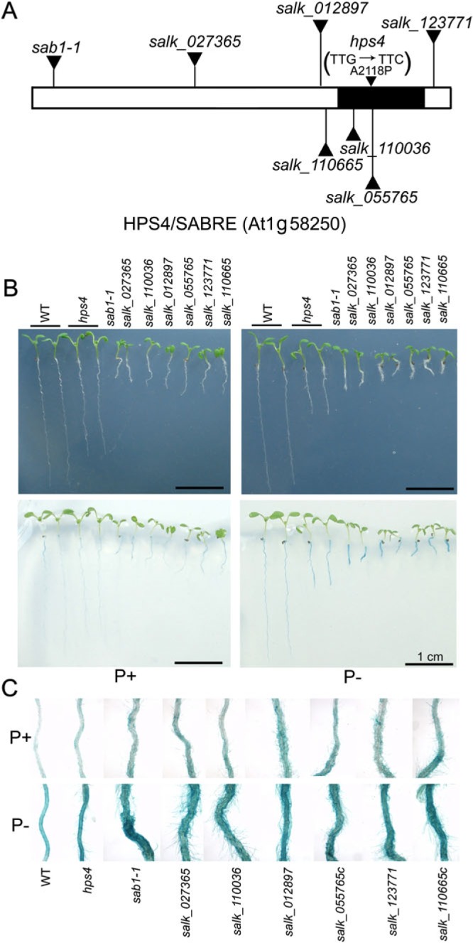
Molecular cloning of the HPS4 gene. (A) A diagram of the structure of the HPS4/SABRE protein. The AGI code of the HPS4 gene is indicated. The filled region indicates the segment that shares sequence homology with a group of Golgi-localized plant proteins. The positions of T-DNA insertions in the sab1-1mutant line and six SALK lines, and the position of the point mutation in hps4 are indicated. The changes in nucleotide and amino acid in the hps4 mutant are shown in parentheses. (B) Morphologies and BCIP staining of 9-day-old seedlings of the WT, hps4, sab1-1, and six SALK T-DNA insertion lines grown on P+ and P– medium. (C) Close-up view of BCIP staining of the seedlings shown in B.