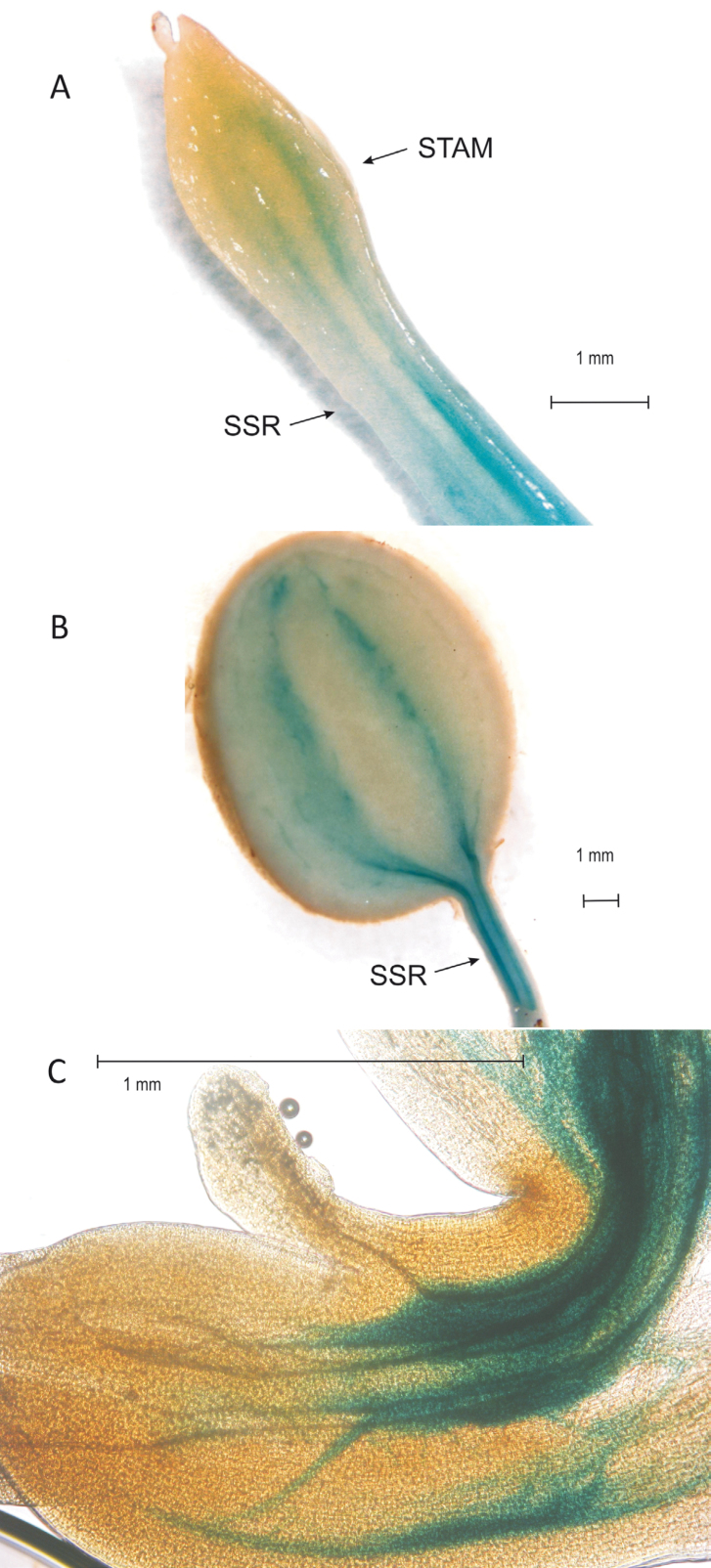
Fig. 2.
Staining of DR5::GUS in transgenic stolon after 9 d in SDs (A) and in a young tuber after 20 d in SDs (B). The arrows indicate the subswelling region (SSR) and the stolon apical meristem (STAM). In (C), staining of promPINV::GUS transgenic plants in the stolon apical hook under LD conditions is shown. The GUS staining is located around the vascular tissue in the STAM, an indication of auxin transport from the site of biosynthesis in the STAM to the basal parts of the stolon. (This figure is available in colour at JXB online.)

