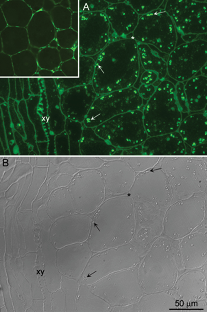
Fig. 8.
Immunodetection of HvPap-1 protease in barley embryos after 24 h of water imbibition. (A) Immunofluorescence localization of HvPap-1 on 2 μm sections of formaldehyde-fixed and LR white-embedded barley embryos 24 hai. Numerous bright foci of different sizes (arrows) are observed within the cytoplasm, excluding the vascular bundle (xy). (B) The fluorescent foci correspond to refringent structures (arrows) revealed by differential interference contrast (DIC). The cell walls (asterisk), clearly identified by DIC, also show some unspecific autofluorescence as seen in the negative control (inset in A).

