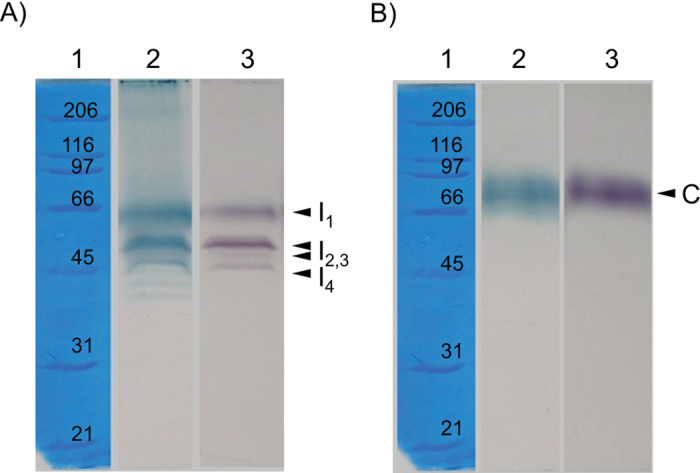
Fig. 1.
Separation of peroxidase isoforms from ionic (A) and covalent (B) cell wall fractions by modified SDS–PAGE. Lane 1, protein standards with their corresponding molecular weight. Gels were stained for haem with TMB (lane 2) and with α-chloro-naphthol for peroxidase activity (lane 3). Arrows indicate different peroxidase isoforms. Four ionic peroxidases isoforms (I1–I4) and one covalent isoform (C) were identified.

