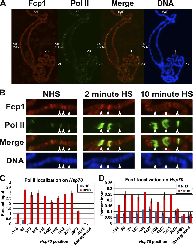
Fig 1.
Fcp1 localizes to transcriptionally active loci. (A and B) Drosophila spread polytene chromosomes immunostained with antibodies to Fcp1 (red) and serine 5-phosphorylated Pol II CTD (H14 antibody, green). The DNA is stained with 4′,6′-diamidino-2-phenylindole (blue). Merge is an overly of Fcp1 and serine 5-phosphorylated Pol II CTD. Panel A shows chromosomes from salivary glands under NHS conditions. In panel B, Hsp70 loci (87A and 87C [endogenous] and a single Hsp70 transgene at 87E) are marked by arrows in salivary glands under NHS and HS conditions. (C) ChIP results showing the enrichment of Pol II (Rpb3) at the Hsp70 gene in Drosophila S2 cells under NHS and HS conditions. (D) ChIP results of the Fcp1 enrichment on the Hsp70 gene in Drosophila S2 cells under NHS and HS conditions. The x axis shows the midpoint of each PCR fragment along the Hsp70 gene, and the y axis shows the percentage of input DNA immunoprecipitated (error bars indicate the standard error of the mean of at least four biological replicates).

