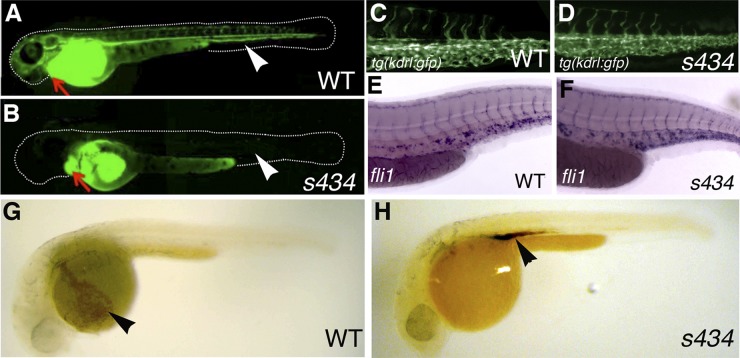
Fig 2.
s434 mutants do not demonstrate blood circulation; however, they do possess intact vasculature. (A and B) Microangiography was performed at 48 hpf with fluorescein-dextran, injected into the sinus venosus, just posterior to the atrium. An arrow is pointing to the heart in both images. In the WT (A), blood flow carries the labeled dextran out into the distal vessels of the embryo, visualized in green. Arrowheads denote the locations of the dorsal aorta and posterior cardinal vein, which can be used to assess circulation. In the s434 mutants (B), however, the dextran does not circulate throughout the embryo. (C and D) kdrl:GFP expression at 48 hpf in both WT (C) and mutant (D) embryos in the trunk, displaying intact endothelial cells in the vessels. (E and F) In situ hybridization for endothelial marker fli1 expression in the trunk region of WT (E) and mutant (F) embryos at 48 hpf. (G and H) o-Dianisidine heme staining at 33 hpf. The wild-type (G) embryo displays circulation, and broad heme staining is observed in the blood cells within the common cardinal vein (ccv) (arrowhead). The mutant (H), however, does not demonstrate circulation (lack of staining in the ccv), and blood cells are present in the intermediate cell mass region above the yolk extension (arrowhead), at the site of their formation.

