Fig 5.
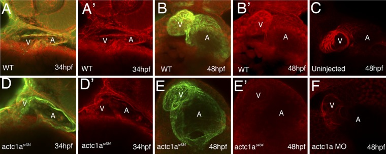
The actc1as434 mutation results in a dramatic decrease in the amount of polymerized F-actin present in the heart. F-actin was stained with rhodamine-phalloidin (red channel; panels A, A′, B, B′, D, D′, E, and E′), and an MF20 antibody stained for a sarcomeric myosin (green channel). (The green channel is overlaid with the red channel in panels A, B, D, and E.) In the ventral view, the anterior portion is up; maximum-intensity projections of confocal images are shown. At 34 hpf, the F-actin staining in the mutant (D and D′) is reduced in the heart compared to that in the wild type (A and A′). At 48 hpf, the wild-type heart shows strong staining for both MF20 and F-actin staining, which is particularly pronounced in the ventricle (B and B′). In contrast, the mutant shows a dilated heart and unchanged MF20 but vastly decreased actin staining (E and E′). (C and F) Actc1a-morpholino-oligomer-injected embryos (F) demonstrate a dilated heart tube and decreased F-actin staining compared to the uninjected embryo (C). A, atrium; V, ventricle.

